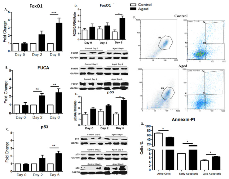
Figure 4.
(A–C) Effects of myoblast aging on the expression of pro-apoptotic genes during myogenic differentiation. Quantitative analysis of FoxO1 (A), FUCA (B), and p53 (C) mRNA expression in aged myoblasts compared to controls during their differentiation. The mRNA expression values of pro-apoptotic factors in the aged myoblasts have been normalized to the corresponding GAPDH mRNA and are expressed as fold changes compared to control myoblasts. (D,E) Effects of myoblast aging on the expression of pro-apoptotic proteins FoxO1 and p53.Representative Western blots and immunoblotting quantification of FoxO1 (D) and p53 (E) in aged myoblasts compared to control cells during their myogenic differentiation. The values of the apoptotic proteins were normalized to each corresponding GAPDH on the same immunoblot. (F,G) Effects of myoblast aging on cell death (Annexin-PI). (F) Histograms from a representative experiment show the apoptotic effect of senescence on myoblasts. The percentages of necrotic, live, early apoptotic, and late apoptotic cells are displayed in R1, R2, R3 and R4, respectively. (G) Bar graphs show that senescence induced the apoptosis of (aged) myoblasts. Quantitative results (R2–R4) are displayed aspercentagechanges compared to the control. (Mean ± SE of 3 independent experiments performed in triplicate; * p < 0.05, ** p < 0.01, *** p < 0.001).

