Figure 1.
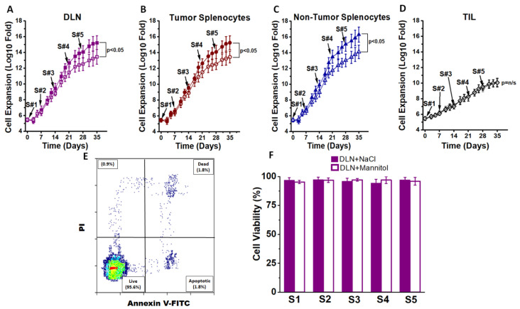
Ex vivo expansion of CD4+T cells. (A) The CD4+T cells obtained from draining lymph nodes (DLNs) of day 28 Py230-othrotopic syngeneic breast tumor bearing mice were expanded in the presence of high salt culture treatment (∆0.035 M NaCl, filled boxes) or with control equimolar mannitol (0.035 M mannitol, open boxes). CD4+T cells were subjected to five stimulation cycles (S1–S5), with each cycle lasting for 7 days and a total of 35 days. Stimulation cycle details including other chemicals used in the cell culture are provided in the “Materials and Methods” and “Results” sections. (B) The CD4+T cells obtained from the spleens of day 28 tumor bearing mice were expanded in the presence of high salt culture treatment (filled circles) or with control equimolar mannitol (open circles). (C) The CD4+T cells obtained from the spleens of 12-week-old non-tumor bearing mice were expanded in the presence of high salt culture treatment (filled triangles) or with control equimolar mannitol (open triangles). (D) CD4+Tumor infiltrating lymphocytes (TILs) were isolated by magnetic bead separation and stimulated for 5 cycles with either high salt (filled grey diamond) or mannitol (open diamond). (E) Cell viability test for staining with propidium iodide and annexin V staining. (F) Cell viability reported at the end of each 7 day stimulation cycle on CD4+T cells obtained from DLNs. A similar >95% viability was noted on CD4+T cells obtained from tumor splenocytes, non-tumor splenocytes and TILs. Further, no difference in viability was noted between salt treatment and mannitol treatment in any of the four cohorts mentioned above. Data presented as mean ± standard error of mean (SEM) from 5 independent CD4+T-cell expansions.

