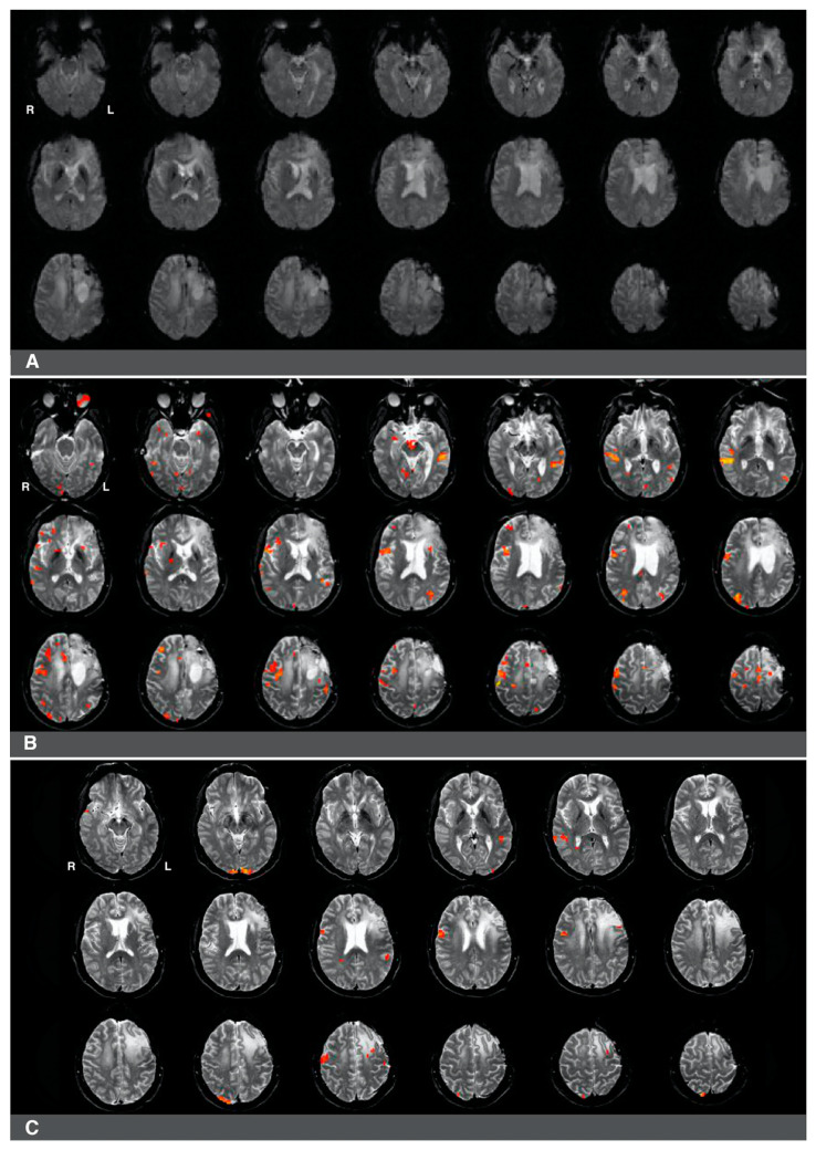
Figure 4.
Right-hemisphere activations in a (clinically confirmed) left language-dominant patient with a high-grade glioma in the left hemisphere. Panel (A): a raw functional image with a notable absence of activation in the left frontal region corresponding to Broca’s area. The absence of activation may be related to artifact and edema from prior resection in the area. Panel (B): Language activations consistent with Broca’s area observed in the right hemisphere during pre-surgical fMRI. Panel (C): Prior fMRI from six months earlier indicates Broca’s representation in the left hemisphere.

