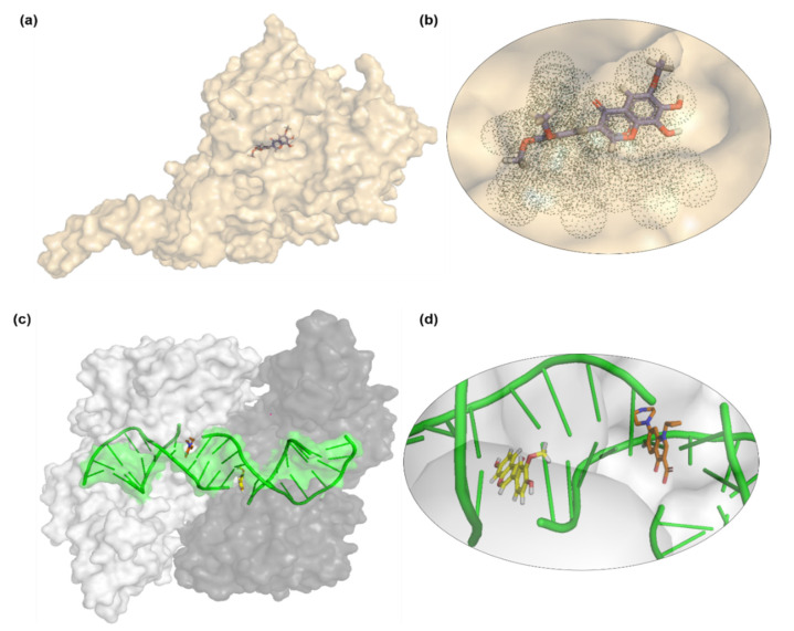
Figure 6.
Binding pocket visualization of (a,b) yeast S. cerevisiae lanosterol 14α-demethylase enzyme (a cytochrome P450 enzyme) docked with compound 7 isolated from Onosma chitralicum. The enzyme is reported in a co-crystallized form with fluconazole under PDB code 4WMZ. Compound 7 (shown in stick form) is visible in the dotted binding pocket, showing that this compound can act as a potential pro-drug and be modified for fitting in the active site groove. (c,d) Docked compound 4 is found in the mirror image form of ciprofloxacin, co-crystallized with S. aureus topoisomerase-II DNA gyrase (PDB ID: 2XCT), involved in hydrogen bonding to the bound DNA molecule. Ciprofloxacin and compound 4 are shown in sticks, bound DNA in green ribbon, and dimeric enzyme in white and black protomers with surface representation.

