Figure 1.
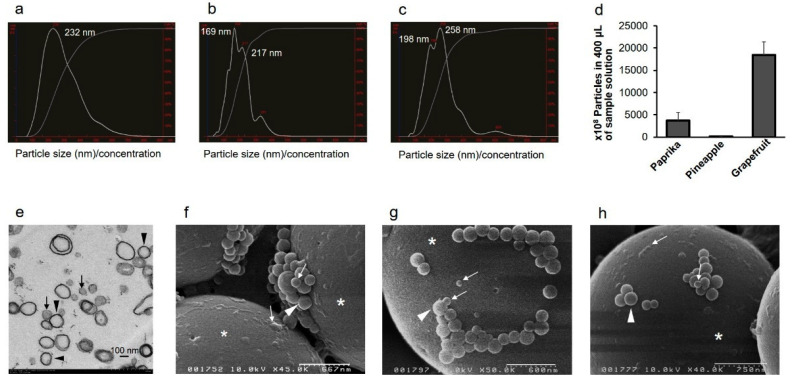
Size characterization of extracellular vesicles (EVs) from fruit and vegetable juice. (a–d) NanoSight analysis of EVs. Representative particle size distributions for EVs are as follows: paprika juice (freshly hand-squeezed juice, SJ) (a), pineapple juice (commercially available 100% natural fruit juice, J) (b), and grapefruit J (c). (d) Number of EVs in juice samples. The data are shown as average ± standard deviation (n = 3). Paprika SJ: 3738 (1815); pineapple J: 105 (15); grapefruit J: 18,467 (2902). (e) Transmission electron microscopy analysis of an EV pellet from grapefruit J. Exosome-like nanovesicles are visible, approximately 50–100 nm (black arrows) and 200 nm (black arrowheads) in diameter. Bar = 100 nm. (f–h) Scanning electron microscopy analysis of EVs. Exosome-like nanovesicles from paprika SJ (f), pineapple J (g), and grapefruit J (h) are shown. The vesicles are approximately 50–100 nm (white arrows) and 200 nm (white arrowheads) in diameter. White asterisks: 3 μm polyethylene beads. Scale bars: f, 667 nm; g, 600 nm; h, 750 nm.

