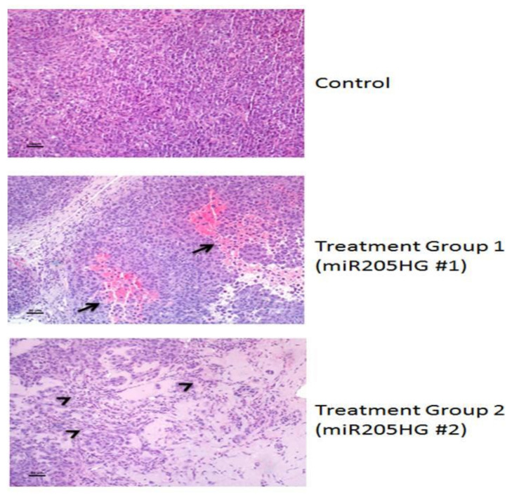
Figure 12.
Hemorrhage, necrosis, leukocyte infiltration, and tumor cell death in miR205HG-treated murine tumors. Xenografts from vector-treated controls were composed of densely packed sheets of cells with only occasional individual cell necrosis and leukocyte infiltration. In contrast, miR205HG-treated xenografts showed extensive areas of hemorrhage and necrosis (arrows) with leukocyte infiltration (miR205HG #1) or areas of tumor cell death, accumulation of proteinaceous fluid, leukocyte infiltration, and necrotic tumor cells (arrowheads) (miR205HG #2). Scale bars = 50 um.

