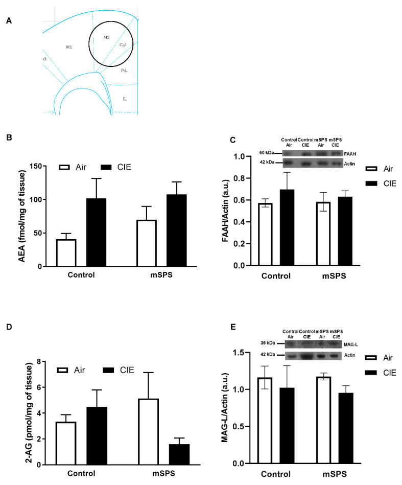
Figure 1.
Anandamide (AEA) and 2-arachidonoylglycerol (2-AG) contents, fatty acid amide hydrolase (FAAH) and monoacylglycerol lipase (MAG-L) levels in the prefrontal cortex (PFC) after mSPS/Control or Air/CIE exposures. (A) Schematic of coronal slice where PFC tissue punches were taken bilaterally (adapted from [58]). Average (B) AEA content (Control-Air: n = 4; Control CIE: n = 7; mSPS-Air: n = 6; mSPS-CIE: n = 8) (C) FAAH levels (Control-Air: n = 6; Control CIE: n = 4; mSPS-Air: n = 5; mSPS-CIE: n = 8) (Inset: representative immunoblotting sample images; kDa kilodaltons) (D) 2-AG content (Control-Air: n = 4; Control CIE: n = 7; mSPS-Air: n = 6; mSPS-CIE: n = 8), and average (E) MAG-L levels (Control-Air: n = 7; Control CIE: n = 4; mSPS-Air: n = 4; mSPS-CIE: n = 7) in the PFC (Inset: representative immunoblotting sample images) did not change among groups after mSPS/Control or Air/CIE exposures. Data are mean ± SEM.

