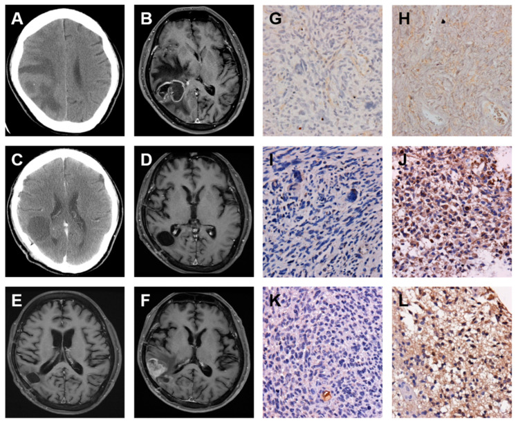
Figure 5.
Enhanced FN, VIM, and TGF-β expressions are associated with the local recurrence of GBM. Serial images including first and follow up examinations of a patient with GBM are shown by using either brain CT on 10 July 2009 (A) and 7 August 2009 (C) or head MRI on 10 July 2009 (B), 30 November 2009 (D), 16 April 2010 (E), and 26 November 2010 (F). The IHC staining images indicate the expression level of FN (G and H), VIM (I and J), and TGF-β (K and L) proteins in origin (G, I, and K) and local recurrence (H, J, and L) of GBM tumor specimens.

