Figure 6.
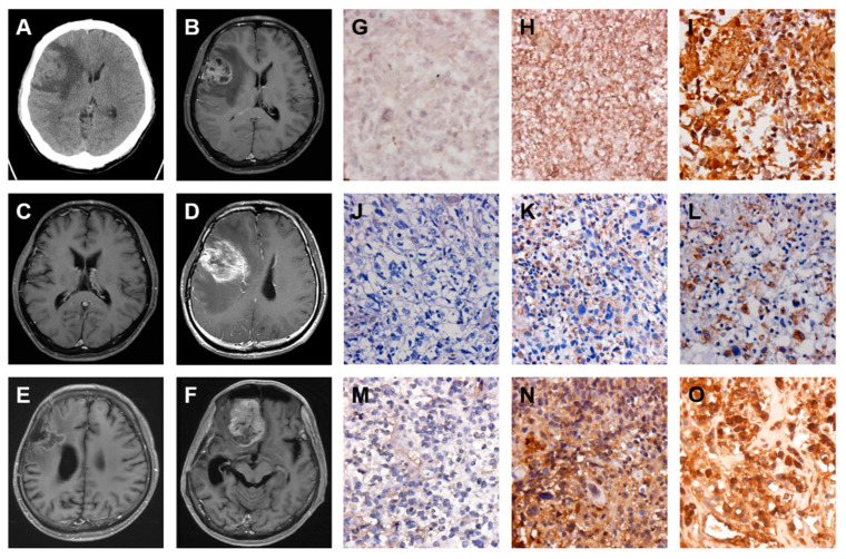
Expression of FN, VIM, and TGF-β is elevated during local recurrence and then remote brain metastasis of GBM. Serial images including first and follow up examinations of a patient with GBM are shown by using either brain CT on 1 August 2010 (A) or head MRI on 1 August 2010 (B), 11 April 2011 (C), 19 July 2012 (D), 30 January 2013 (E) and 3 June 2013 (F). The IHC staining images indicate the expression level of FN (G–I), VIM (J–L), and TGF-β (M–O) proteins in origin (G, J, and M), local recurrence (H, K, and N) and then remote brain metastasis (I, L, and O) of GBM tumor specimens.

