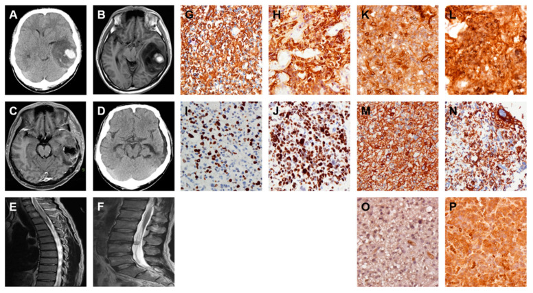
Figure 7.
Increased FN, VIM, and TGF-β expressions are associated with spinal metastasis of GBM. Serial images including first and follow-up examinations of a patient with GBM are shown by using either brain CT on 11 May 2013 (A) and 17 August 2014 (D) or head MRI on 11 May 2013 (B), 26 July 2013 (C) and 17 August 2014 (E and F). The IHC staining images indicate the expression level of glial fibrillary acidic protein (GFAP) (G and H), Ki-67 (I and J), FN (K and L), VIM (M and N), and TGF-β (O and P) proteins in origin (G, I, K, M, and O) and spine metastasis (H, J, L, N, and P) of GBM tumor specimens.

