Figure 6.
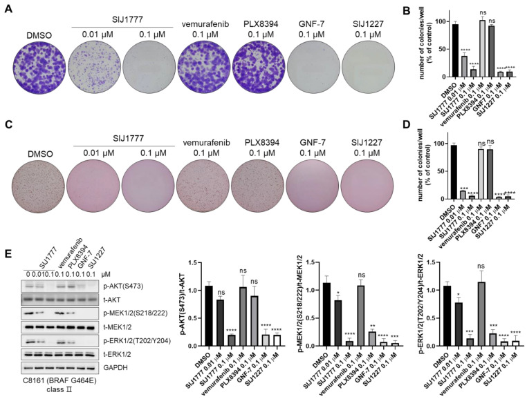
Clonogenic assay analysis of SIJ1777 in C8161. (A,B) 2D clonogenic assay (colony formation assay) results of the compounds on C8161 melanoma cell. After incubation with test compounds for 14 days, colonies were photographed without magnification. (C,D) 3D clonogenic assay (soft agar assay) results of test compounds on C8161 melanoma cell. Cells embedded within 0.35% low melting agar and incubated with the indicated compounds for 14 days and observed without magnification. (B,D) Number of colonies were determined automatically by ImageJ (n = 3, respectively). (E) Western blot analysis of SIJ1777 in C8161. Cells were treated with 0.01, 0.1 μM of SIJ1777, and 0.1 μM of vemurafenib, PLX8394, GNF-7, and SIJ1227 for 24 h. Cell lysates were subjected to western blot analysis to estimate the phospho- or total- form of AKT, MEK, ERK levels, and GAPDH was used as the internal loading controls (left panel). Quantification result (n = 3) of western blot result by ImageJ (right panel). Statistical significances were determined using a one-way ANOVA analysis (* p < 0.05, ** p < 0.01, *** p < 0.001, **** p < 0.0001).

