Figure 1.
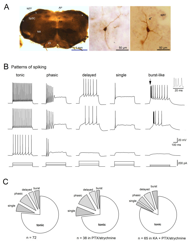
Firing patterns of Sp5C laminae I/II cells. (A) Bright field micrographs illustrate a typical freshly prepared transverse slice (350 µm thickness; left), and examples of biocytin-filled neurons in lamina II (middle) and in lamina I (right, with soma location indicated by an asterisk on the left, and axon pointed by arrow). AP, area postrema; cc, central canal; IO, inferior olivary complex; NA, nucleus ambiguous; sptV, spinal tract of the trigeminal nerve; XII, hypoglossal nucleus. (B) Five classes of Sp5C laminae I/II cells are distinguished by their properties of action potential discharge to depolarizing currents. Cells were recorded under whole-cell current-clamp mode, with membrane potential initially held at −70 mV (by current injection). Pattern of spiking was determined by their responses (top three panels) to the serial of current injection (bottom panels). The initial burst-like spiking (arrow) was enlarged on the right. (C) Distribution of spiking patterns of Sp5C cells recorded in the absence and in the presence of inhibitory and excitatory synaptic blockers. Kynurenic acid (KA, 2 mM) was used to block fast glutamatergic synaptic transmission, and picrotoxin (PTX, 100 µM) plus strychnine (10 µM) were used to block fast GABAergic and glycinergic synaptic transmissions, respectively.

