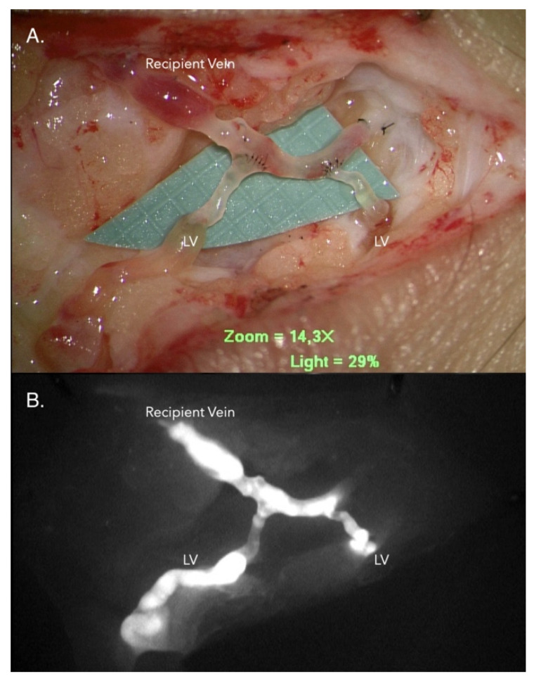
Figure 1.
Lymphaticovenous anastomosis (LVA). (A) Two lymphatic vessels with diameter 0.7 mm and 0.5 mm were anastomosed to a recipient vein in an end-to-side orientation with 11-0 nylon suture. (B) The lumen of the recipient vein became enhanced under near-infrared lymphography. The indocyanine green (ICG)-containing lymph has flowed from the lymphatic vessel into the recipient vein, indicating good antegrade lymphatic flow. Note: the background grid is 1 * 1 mm2.

