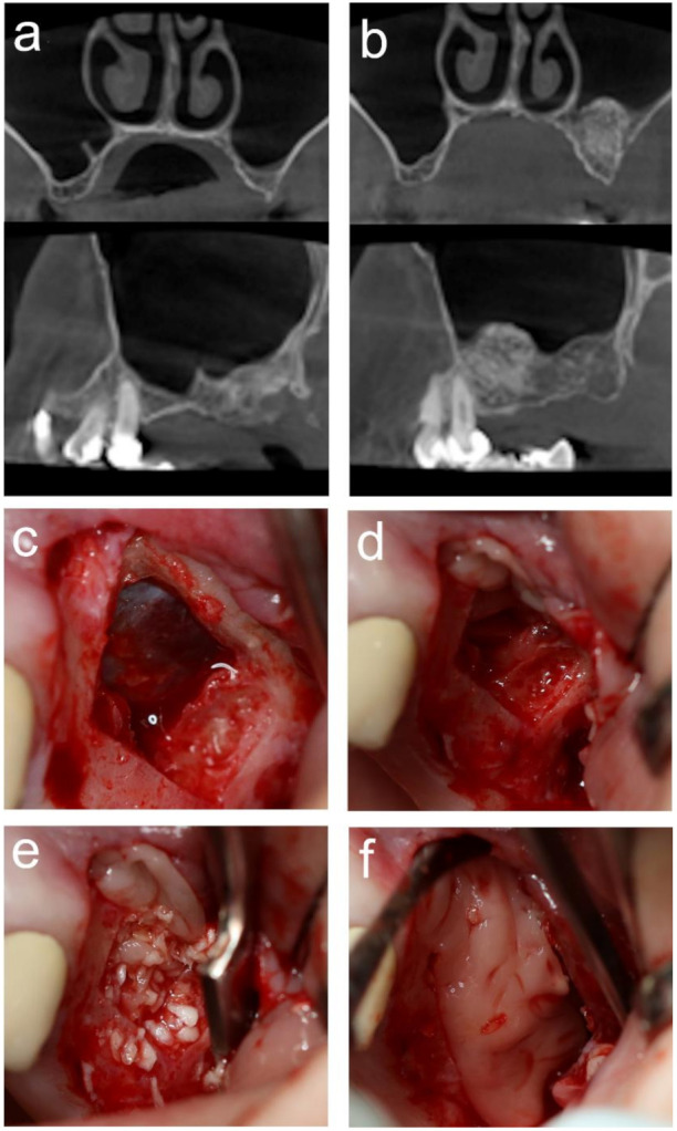
Figure 1.

Representative cone-beam computed tomography (CBCT) images are shown from the test group before maxillary sinus augmentation. (MSA) (a) and after 3 months of healing (b). The main steps of MSA were as follows: piezoelectric osteotomy was carried out, and the bony window was removed to expose the Schneiderian membrane (SM) (c). SM was elevated and then covered with two pieces of advanced platelet-rich fibrin (A-PRF) membrane (d). Serum albumin-coated bone allograft particles were mixed with A-PRF and gently packed into the created space (e). The lateral window was covered with the previously removed bony wall and an A-PRF membrane (f).
