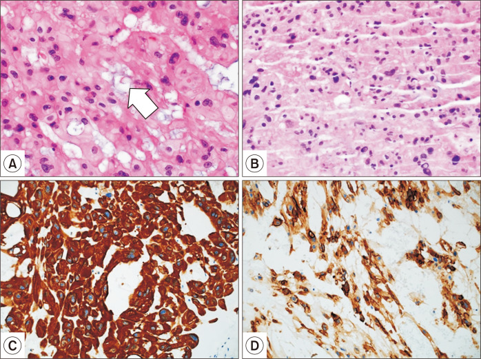
Fig. 2.
(A) Area of tumor populated by large cells with abundant, bubbly cytoplasm (arrow), so-called physaliferous cells, typical of chordomas (H&E, ×200); (B) endoscopic ultrasound-guided fine needle aspiration biopsy failed to provide a definitive diagnosis (H&E, ×200), although pancytokeratin positivity and mucin-filled vacuoles suggested adenocarcinoma (H&E, ×200); (C) pancytokeratin positivity; and (D) epithelial membrane antigen positivity.

