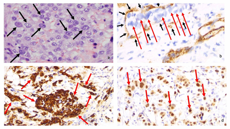
Figure 3. Microscopic examination of cutaneous metastatic salivary duct carcinoma.
Microscopic examination at high magnification of the neoplastic salivary duct carcinoma cells shows large, irregular nuclei (black arrows) with visible nucleoli (a). Immunohistochemistry (brown staining) with anti-cluster of differentiation 31 (anti-CD31) highlights vascular endothelial cells (black arrows) containing neoplastic cells (red arrows), thereby demonstrating vascular invasion by the metastatic carcinoma (b). Anti-cytokeratin 7 (anti-CK7) and androgen receptor (both brown staining) label tumor cells (red arrows) (c and d, respectively) (a, hematoxylin and eosin, ×400; b, anti-CD31, diaminobenzidine immunoperoxidase and light hematoxylin, x 400; c, anti-CK7, diaminobenzidine immunoperoxidase and light hematoxylin, ×400; anti-androgen receptor, diaminobenzidine immunoperoxidase and light hematoxylin, ×400). The images have not previously been published; however, the details of the pathology of the patient’s skin lesions have previously been described [3-5].

