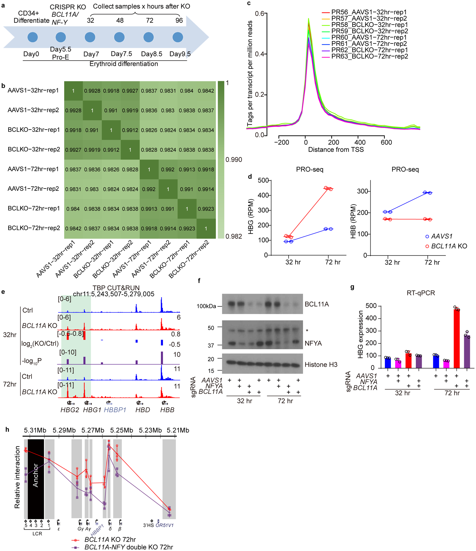
Extended Data Fig. 5. Acute depletion of BCL11A leads to rapid binding of NF-Y.

(a) Schematic diagram of primary human CD34+ differentiation and acute depletion of BCL11A using CRISPR/Cas9.
(b) Pairwise correlation of PRO-seq experiments. All the experiments in each time point showed high degree of correlation, indicating very minor transcriptional fluctuation upon BCL11A depletion.
(c) Average PRO-seq signal at −200 to +600 bp relative to TSS exhibited promoter pausing of PolII.
(d) Quantification of PRO-seq reads on HBG1/2 and HBB genes after 32 or 72 hrs of BCL11A acute depletion. The y-axis shows Reads Per Million (RPM) for HBG1+HBG2 or HBB. The result is shown as mean (SD) of two biologically independent samples (independent cell cultures and CRISPR KO).
(e) CUT&RUN of TBP in CD34+ cells undergoing erythroid differentiation after 32 or 72 hrs of BCL11A acute depletion. The result shown is representative of two biological replicates. Quantification of KO/Ctrl and the corresponding p-values are reported by MAnorm.
(f) Western blot for BCL11A and NFYA in adult primary human CD34+ derived erythroid cells upon KO of NFYA, BCL11A or both (cropped).
(g) RT-qPCR analysis of γ-globin expression in adult primary human CD34+ derived erythroid cells upon KO of NFYA, BCL11A or both. Knockout of NFYA after 72 hours decreases γ-globin expression. The result is shown as mean (SD) of three technical replicates.
(h) Chromosome Conformation Capture qPCR in adult primary human CD34+ derived erythroid cells, comparing BCL11A KO and BCL11A/NF-Y double KO. EcoRI fragment encompassing HS2–4 of the LCR was used as anchor point to evaluate LCR-globin interaction. The result is shown as mean (SD) of three technical replicates.
