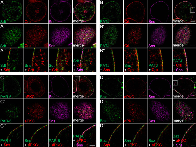
Fig. 1.
Apical polarity regulators localize at the cortex of nephrocytes. A–D Garland nephrocytes were dissected from 3rd instar larvae, fixed and stained with the indicated antibodies. Subsequently, samples were embedded in acrylamide gel and subjected to expansion to increase sample size and thus resolution. Two sections were imaged: the equatorial plane (A–D) and an onview on the surface of the nephrocyte (A′–D′). A″–D″ are magnifications of the cortical region from A–D. Scales bars are 10 µm in A–D, 5 µm in A′–D′ and 3 µm in A″–D″

