Fig. 3.
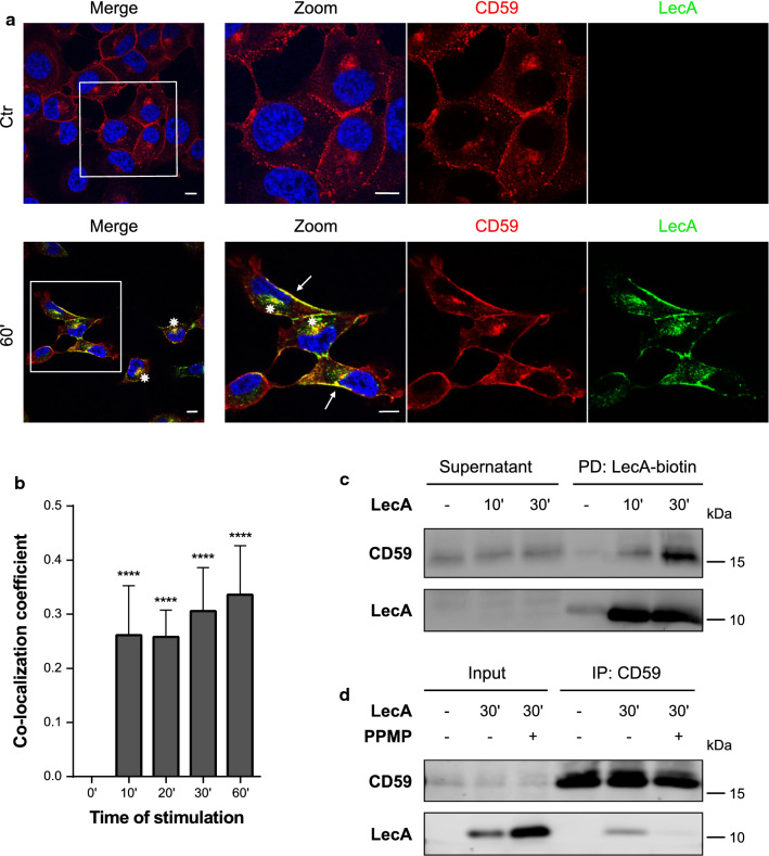
The LecA plasma membrane domain includes CD59 proteins. a Fluorescence co-localization studies of CD59 (red) and LecA (green) after 60 min of lectin stimulation. Nuclei were counterstained by DAPI. Framed areas were magnified. White arrows point at co-localization events at the plasma membrane, asterisks at perinuclear co-localization. Scale bar: 10 μm. b Signal overlay of LecA and CD59 is displayed by Mander’s co-localization coefficient and statistically compared to time point 0. Bars display mean values of at least four biological replicates, error bars represent SD, ****p < 0.0001 (one-way ANOVA and Dunnett’s multiple comparison tests). c CD59 was validated as component of the LecA-binding domain by pull-down of LecA-biotin. d Immunoprecipitation of CD59 co-precipitated LecA after 30 min of stimulation. PPMP treatment to deplete cells in glucosylceramide-based GSLs, such as Gb3, largely inhibited the precipitation of LecA

