Fig. 6.
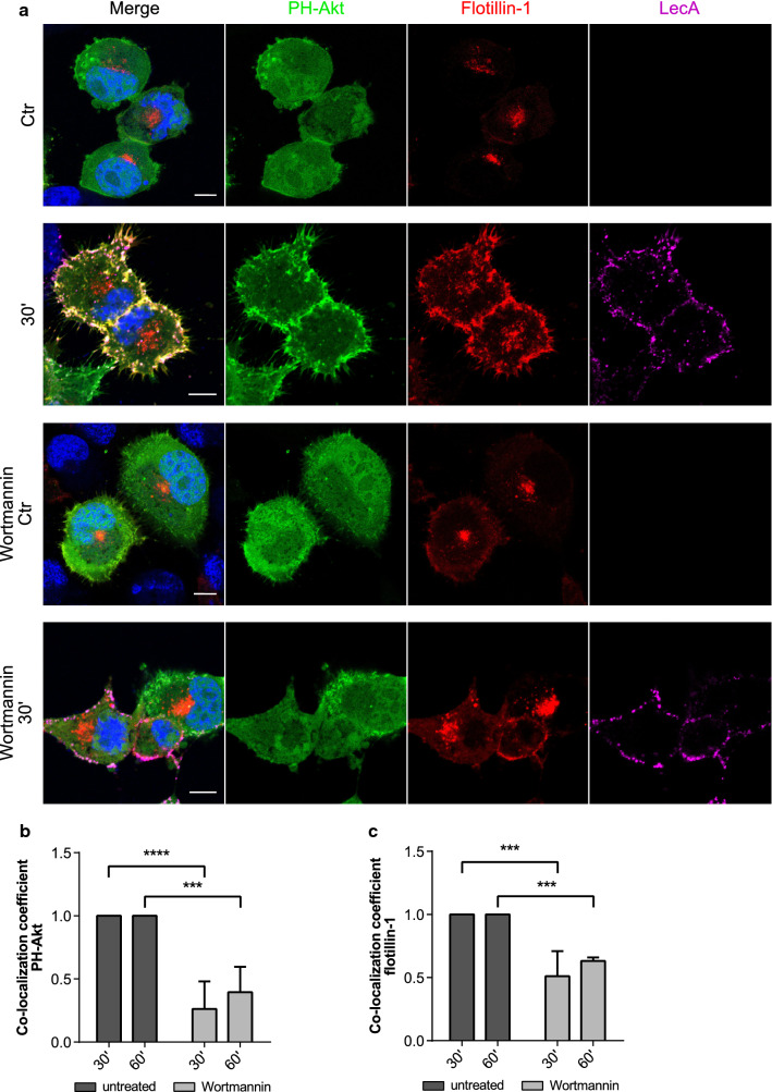
PI3-kinase inhibition diminishes PIP3 clustering and recruitment of flotillins upon LecA stimulation. a Confocal microscopy images of PH-Akt-GFP and flotillin-1-mCherry expressing H1299 cells exposed to fluorescent LecA. Lower two panels: cells were pre-treated with 100 nM Wortmannin to inhibit PI3-kinase activity. Scale bar: 10 μm. b Co-localization of PH-Akt-GFP and LecA is depicted as fold change of Mander’s co-localization coefficient normalized to the untreated conditions. c Fold change of Mander’s co-localization coefficient quantified between the fluorescence signals of flotillin-1-mCherry and LecA in comparison to the untreated conditions. For all panels: bars display mean values of three biological replicates, error bars represent SD **p < 0.01, ***p < 0.001, ****p < 0.0001 (two-way ANOVA and Tukey’s multiple comparisons tests)

