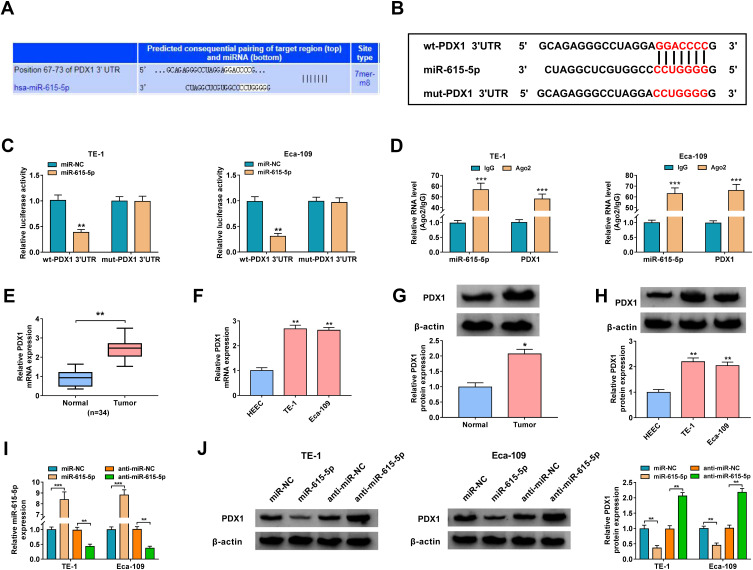
Figure 5.
PDX1 acted as a target of miR-615-5p. (A) The complementary binding sequence between miR-615-5p and PDX1 was predicted by TargetScan. (B) Schematic diagram of the luciferase reporter containing wt-PDX1 3ʹUTR or mut-PDX1 3ʹUTR. (C) Dual-luciferase reporter assay was performed to validate the relationship between miR-615-5p and PDX1. **P < 0.01 vs miR-NC. (D) QRT-PCR presented the enrichment of miR-615-5p and PDX1 in Ago2 and IgG immunoprecipitates. ***P < 0.001 vs IgG. (E–H) Relative expression levels of PDX1 mRNA and protein in ESCC tissues and cells were assessed using qRT-PCR or Western blotting. *P < 0.05 and **P < 0.01 vs para-carcinoma tissues or HEEC cells. (I) Relative expression of miR-615-5p in TE-1 and Eca-109 cells transfected with miR-NC, miR-615-5p, anti-miR-NC, or anti-miR-615-5p was analyzed using qRT-PCR. **P < 0.01 and ***P < 0.001 vs miR-NC or anti-miR-NC. (J) Influence of miR-615-5p overexpression or inhibition on the protein level of PDX1 in TE-1 and Eca-109 cells was evaluated using Western blotting. **P < 0.01 vs miR-NC or anti-miR-NC.

