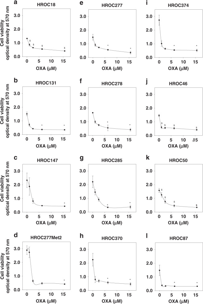
Fig. 1. Oxaliplatin inhibits cell viability.
HROC18 (a), HROC131 T0 M3 (b), HROC147Met (c), HROC277Met2 (d), HROC277 T0 M1 (e), HROC278Met T2 M2 (f), HROC285 T0 M2 (g), HROC370 (h), HROC374 (i), HROC46 T0 M1 (j), HROC50 T1 M5 (k) and HROC87 T0 M2 (l) were treated with appropriate vehicle, 1 µM, 2.5 µM, 6.25 µM or 15.6 µM oxaliplatin (OXA) for 5 days. Oxaliplatin significantly inhibited the cell viability. $ indicates P < 0.001, which was determined by one-way analysis of variance with Holm–Sidak’s post hoc test; * indicates P < 0.05, which was determined by Kruskal–Wallis one-way analysis of variance on Ranks with Tukey’s post hoc test. For (e), (g), (h) and (i), N = 6; for the other cell lines, N = 4.

