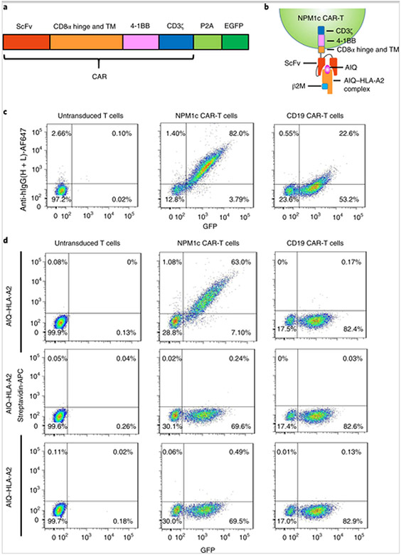
Fig. 3. Generation of NPM1c CAR-T cells specific to the AIQ–HLA-A2 complex.
a, Schematic of the CAR vector consisting of scFv (YG1 or CD19), the CD8α extracellular hinge and transmembrane domain (TM), the 4-1BB co-stimulatory domain and the CD3ζ activation domain, followed by self-cleavage P2A and EGFP. b, Schematic of the recognition of NPM1c CAR-T cells by the soluble AIQ–HLA-A2 complex. c, Flow cytometry analysis of CAR expression by untransduced and transduced T cells. Transduced T cells were enriched by sorting for GFP+ cells, expanded and stained with AF647-labelled antibody specific for human IgG heavy and light chains. Untransduced T cells were activated and expanded without sorting. Shown are the GFP versus anti-human IgG staining profiles of live cells (DAPI−). d, NPM1c CAR-T cells recognize the AIQ–HLA-A2 complex. Untransduced and transduced T cells were incubated with biotinylated AIQ–HLA-A2, SLL–HLA-A2 or HLA-A2, followed by streptavidin-APC staining. Shown are the GFP versus streptavidin-APC staining profiles of live (DAPI−) untransduced T cells, NPM1c CAR-T cells and CD19 CAR-T cells. The data in c and d are representative of three separate experiments. The percentages indicate the percentages of cells in the gated regions.

