
Figure 2.
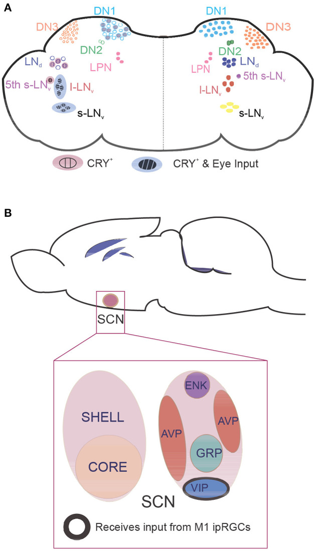
Location of clock neurons in fly and mouse brains. Neurons that express core clock proteins localize in specific regions of the cortex. (A) In flies, ~150 clock neurons can be generally categorized as dorsal neurons (DNs) or lateral neurons (LNs). The right hemisphere contains clock neuron populations, and the left hemisphere shows whether these neurons receive retinal input and/or are intrinsically photosensitive due to CRY expression. (B) In mammals, core clock neurons are found in the suprachiasmatic nucleus (SCN). Neurons in the photosensitive SCN core primarily express the neuropeptides vasoactive intestinal polypeptide (VIP) and gastrin-releasing peptide (GRP), whereas neurons in the SCN shell express arginine vasopressin (AVP) and enkephalin (ENK). M1 intrinsically photosensitive retinal ganglion cells innervate VIP neurons to pass photic information that synchronizes the clock. CRY, cryptochrome; l-LNv, large ventrolateral neuron; LNd, dorsal lateral neuron; LPN, lateral posterior neuron; s-LNv, small ventrolateral neuron.