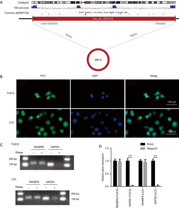
Figure 3.
The characteristics of circ-RANBP9. (A) The ideogram of genomic location and splicing mode of circ-RANBP9. (B) Fluorescence in situ hybridization (FISH) analysis for circ-RANBP9 was performed in TU212 and LCC cells. Scale bar: 100 µm. (C,D) The RNA enzyme digestion test was conducted to confirm the stability of circ-RANBP9. Data indicate mean ± SD, n=3; ***, P<0.001.

