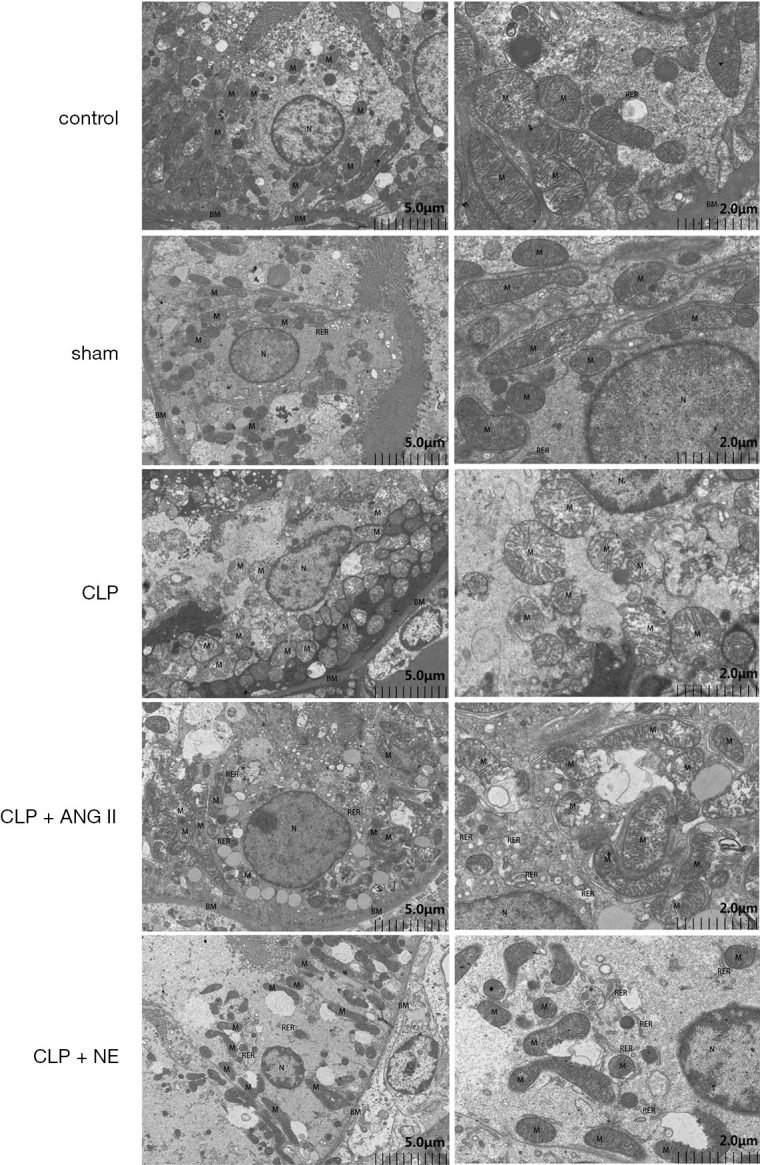
Figure 6.

Effects of ANG II and NE on mitochondrial impairment. Rats were euthanized 24 h after the experiment commenced. A transmission electron microscopy analysis was performed to assess mitochondrial impairment in rat renal cortex tissue samples. Representative images are shown. control and sham group: proximal tubular epithelial cells, with abundant cytoplasmic matrix, continuous and complete basement membrane (BM), uniform thickness and relatively normal structure. The mitochondria (M) were round or rod-shaped, with normal size, large number, clear structure, clear cristae and well-arranged matrix. CLP: epithelial cells of proximal tubules, with severe cytoplasmic edema, obvious weakening of intracellular matrix, local hyperplasia and multilayered basement membrane (BM). Mitochondria (M) swelled and became larger and rounder, more mitochondria cristae arranged irregularly, some mitochondria membrane damaged, inner cristae ruptured, matrix became shallower, and a few mitochondria appeared vacuolar degeneration. CLP + ANG II group: proximal tubular epithelial cells showed moderate cytoplasmic edema, slightly lighter intracellular matrix, more vacuoles, and continuous and complete basement membrane (BM). Mitochondrion (M) swelled moderately, became larger and rounder, most mitochondria swelled, internal cristae became shorter and less, matrix became shallower, a few severe membranes damaged, matrix overflow. CLP + NE group: proximal tubular epithelial cells showed moderate cytoplasmic edema, a large area of low electron density edema area, few organelles, and continuous and complete basement membrane (BM). Mitochondrion (M) was moderately swollen, most of the mitochondria swelled locally, the internal cristae became shorter and less, and the matrix became shallower. In a few area, the membrane damaged and the matrix overflowed. BM: basement membrane. M: mitochondrion. N: nucleus. RER: rough endoplasmic reticulum. Scale bar = 2 µm, 5 µm.
