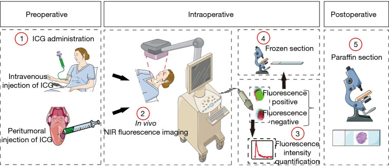
Figure 2.
Workflow of NIR fluorescence imaging for patients with HNSCC. Patients were enrolled in two cohorts: intravenous injection of ICG and peritumoral injection of ICG. After ICG was given, NIR fluorescence imaging was performed in the cervical region, and fluorescence-positive LNs were sent for frozen section intraoperatively. After the fluorescence intensity was recorded, all LNs were sent to undergo paraffin section. NIR, near-infrared; HNSCC, head and neck squamous cell carcinoma; ICG, indocyanine green; LNs, lymph nodes.

