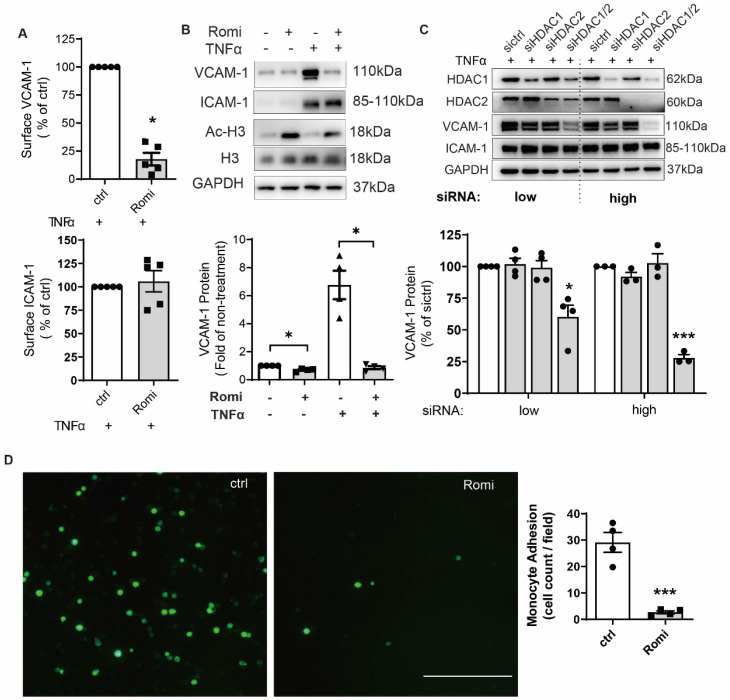
Figure 1.
Inhibition of HDAC1/2 attenuated TNFα-induced VCAM-1 surface expression on HAEC and subsequent monocyte adhesion. (A-B) HAEC were treated with 40 nM Romidepsin (Romi) for 1 h before incubation with 0.1 ng/mL TNFα for 4 h followed by flow cytometry (n = 3, A) and Western blot analysis (n = 4, B) of VCAM-1 and ICAM-1 expression. Increase in the acetylation level of HDAC substrate Histone H3 (Ac-H3) confirms the activity of Romidepsin. Shown below is quantification of VCAM-1 protein (C) HAEC were transfected with 20 nM (low) or 50 nM (high) control siRNA (sictrl), or siRNA targeting HDAC1 (siHDAC1), HDAC2 (siHDAC2) or both (siHDAC1/2) before stimulation with 0.1 ng/mL TNFα for 4 h followed by Western blot analysis. VCAM-1 expression was quantified with Image J (n = 4). (D) HAEC were pretreated with Romi followed by stimulation with TNFα as described above. Adhered DiO-stained THP-1 cells were visualized by fluorescence microscopy and quantified (n = 4). Scale = 100 μm. *p < 0.05; ***p < 0.001, one sample t-test (A, B); paired two-tailed t-test (D); repeated measures one-way ANOVA followed by Dunnett's test (vs. sictrl, C).

