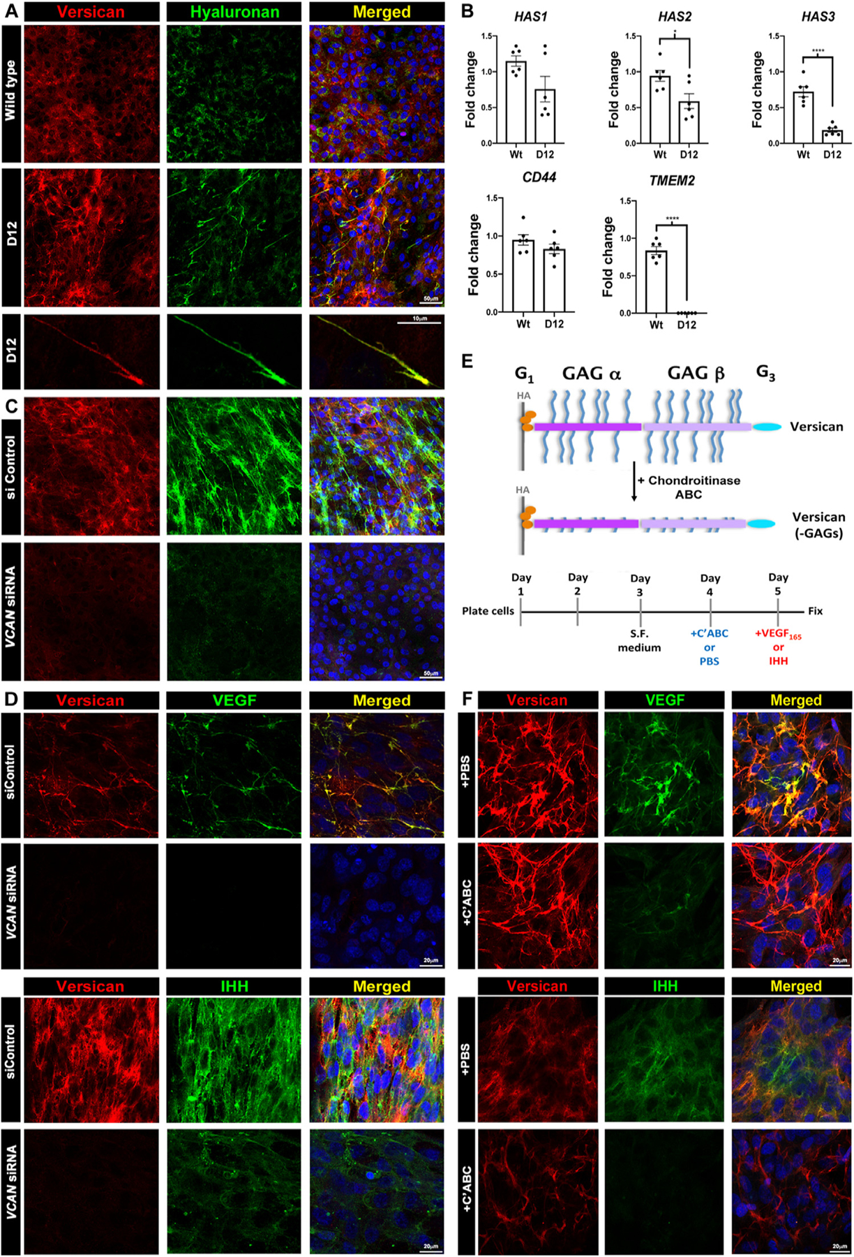
Fig. 8.

Versican regulates HA abundance and cable formation and sequesters VEGF and Ihh via chondroitin sulfate (CS) chains in a cell culture model. (A) Increased versican (red) and HAbp staining (green) in ADAMTS9-deficient (D12) RPE-1 cells. Versican and HA co-stained cables are formed in D12 cells. (B) RT-qPCR showing reduced HAS2 and HAS3 mRNA but not HAS1 or CD44 and dramatically reduced TMEM2 expression in D12 cells. (n=3 independent RNA extractions, error bars= S.E.M., *, p<0.05; ****, p<0.0001). (C) VCAN knockdown reduces both versican (red) and HA staining in D12 cells. (D) Recombinant VEGF165 or Ihh (green) co-staining with versican (red) in control siRNA-transfected D12 cells is lost in VCAN siRNA-transfected D12 cells. (E) The schematic illustrates chondroitinase ABC removal of versican GAG chains and the experimental timeline used. (F) Fluorescence microscope of VEGF and Ihh with versican, with or without chondroitinase ABC treatment prior to addition of recombinant VEGF165 or Ihh (green) shows reduced binding of both growth factors after CS-chain removal, which does not affect versican core protein staining (red). Scale bars in A are 50μm and 10μm, 20μm in D and F and 50μm in C.
