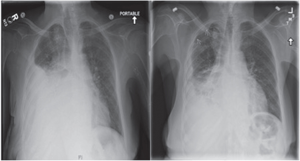
FIGURE 1.
Pneumothorax ex vacuo. This anterior-posterior chest radiograph (left) demonstrates a large pleural effusion in a patient with a chronic exudative right-sided effusion. After thoracentesis, repeat posterior-anterior chest radiography (right) showed a partially re-expanded lung with persistent effusion and evidence of apical pneumothorax, as indicated by arrows, consistent with pneumothorax ex vacuo.

