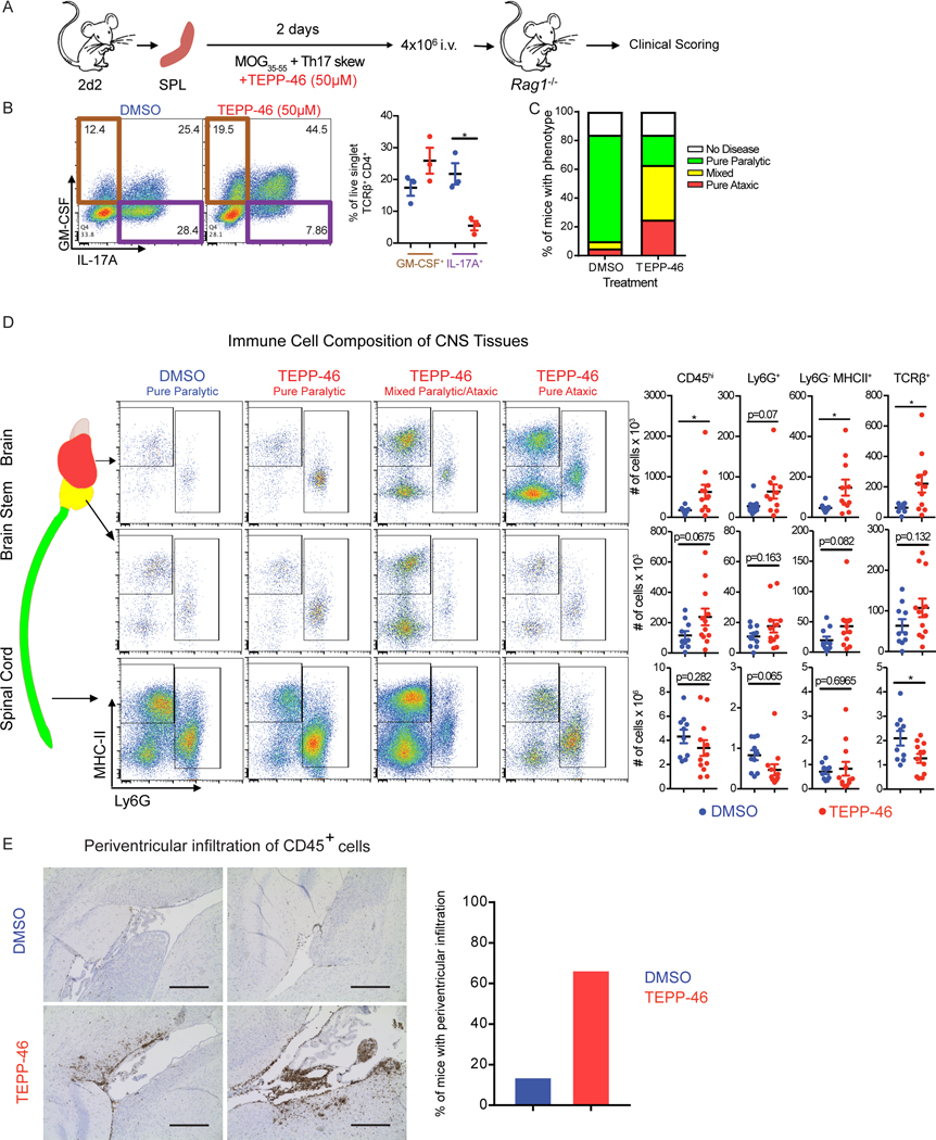
Fig. 7: PKM2 activator treatment redirects TH17 cells to adopt an encephalitogenic program.
(A) Schematic showing the stimulation of splenocytes from 2D2 mice and passive EAE induction. (B) Flow cytometry analysis of IL-17A and GM-CSF production by TCRβ+CD4+ cells on the day of splenocyte transfer. *p<0.05 by repeated measures one-way ANOVA with Tukey post test. (C) EAE phenotypes observed at day 16 post transfer of splenocytes treated with TEPP-46 (50μM) or vehicle (DMSO). Data shown are from n=19 DMSO- and n=24 TEPP-46-treated Rag1−/− mice that received 2D2 cells, pooled from four independent experiments. (D) Flow cytometry analysis of MHC-II and Ly6G expression on myeloid cells (defined as live, singlet, CD45hiCD11b+ cells) isolated from the indicated parts of the CNS of Rag1−/− mice receiving DMSO- or TEPP-46-treated cells. Data are expressed as mean ± SEM from n=10–12 mice per group pooled from N=2 independent experiments. Two-tailed Welch’s t-test. *p<0.05. (E) Representative images (left panels) of anti-CD45 staining in the periventricular brain regions from recipients of DMSO- or TEPP-46-treated splenocytes and quantification (right panel) or mice presenting with perivascular infiltrates. Data are compiled from two independent experiments with a total of 6 mice. Scale bar is 400 μm.

