Figure 1.
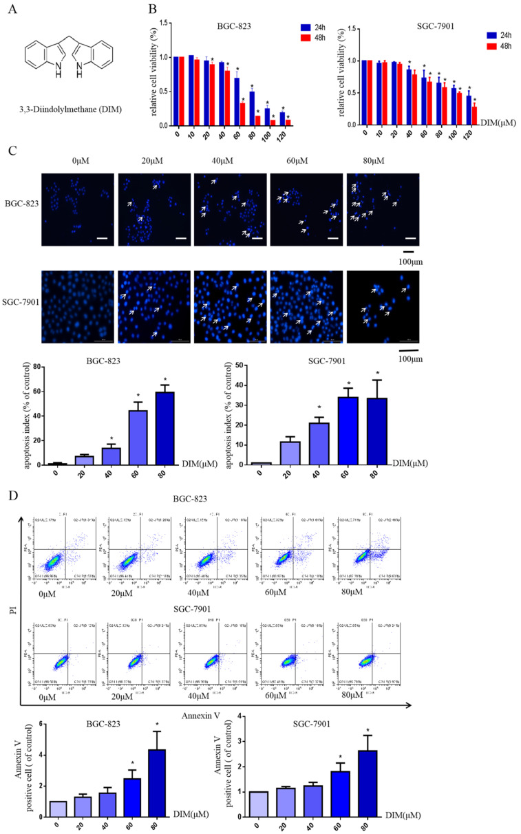
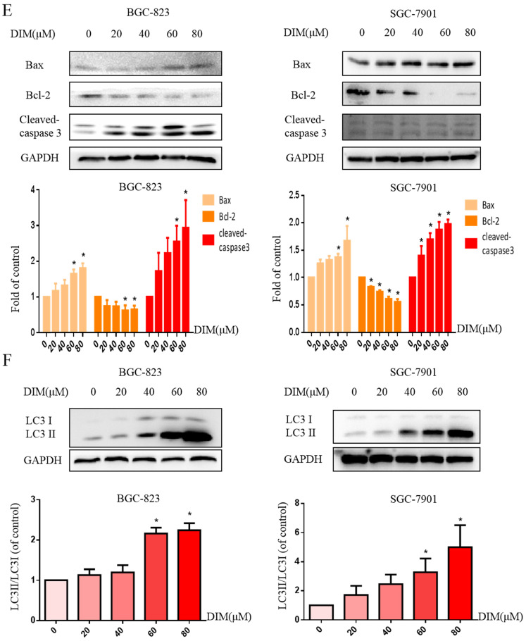
DIM reduces cell viability and induces autophagy and apoptosis in gastric cancer. (A) The structural component of DIM. (B) Cells were treated with different DIM (0-120μM) for 24, 48h, which viability was determined by MTT assay. The results are presented as mean±SD and described as column chart *p<0.05 as compared with control group. (C) Hoechst 33342 staining was used to evaluate apoptosis levels with treatment of DIM for 24h. (D) Flow cytometry was assessed cells apoptosis with DIM treatment. (E) Cells apoptotic proteins analysis of DIM treated cells. BGC-823 and SGC-7901 gastric cancer cells treated with DIM (0-80μM) for 24h. The expression of cell key apoptosis-related protein was detected by Western-blotting. DIM significantly increase the pro-apoptotic marker Bax, cleaved-caspase 3 and decrease anti-apoptotic marker Bcl-2 compared control group. (F) Cells exposure with DIM (0-80μM) for 24h, the expression of autophagy-related proteins was detected by Western-blotting. DIM significantly increase LC3II/LC3I. Values represent as the mean±SD of three independent experiments (n=3) (*p<0.05 compared with the control group).


