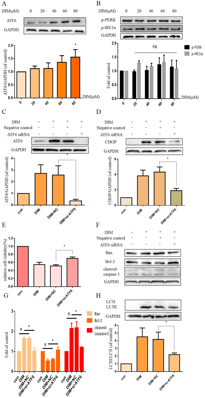
Figure 3.
DIM triggers the activation of UPR. (A, B) BGC-823 gastric cancer cells were treated with 80μM DIM for 24h. The levels of p-PERK, ATF6, p-IRE1α were analyzed by western blot. Quantitative analysis of protein levels. Values are expressed as the mean±SD of three independent experiments (n=3) (*p<0.05 compared with the control group). (C) The knockdown efficiency of ATF6 was evaluated by western blot in cells transfected with negative control siRNA or ATF6 siRNA. (D, F, H). The protein levels of CHOP, Bax, Bcl-2, cleaved-caspase 3, LC3II/LC3I was detected by Western blot in cells transfected with NC siRNA or CHOP siRNA. (E) Cells were transfected with NV siRNA or ATF6 siRNA for 48h, followed by incubation with 80μM DIM for another 24h, cells viability was measured by the MTT assay. The results are presented as mean±SD and described as column chart *p<0.05 as compared with control group. (G) Quantitative analysis of protein levels. (Values represent as the mean±SD of three independent experiments (n=3)(*p<0.05 compared with the control group)).

