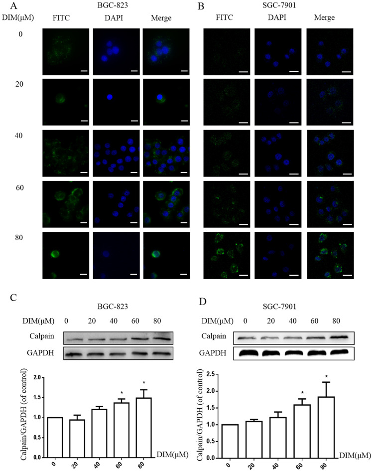
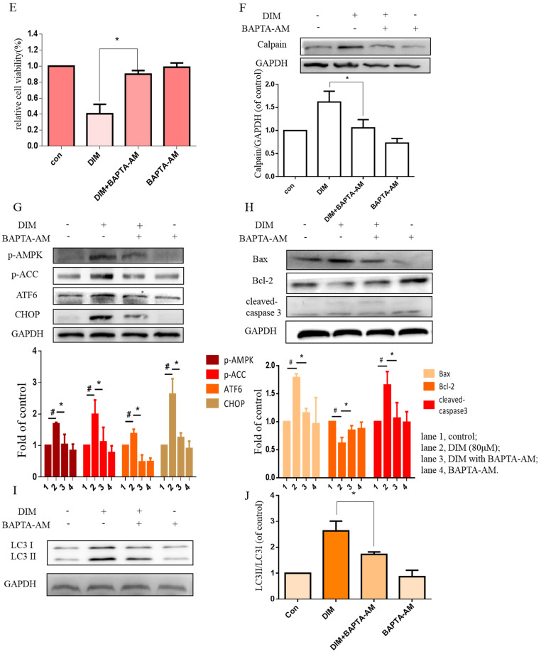
Figure 5.
Cytoplasmic free calcium was involved in DIM-induced cells death by p-AMPK-mediated ER stress. (A) The human BGC-823 gastric cancer cells were treated with different concentration of DIM for 24h, fluorescence intensity of Ca2+ was measured by laser scanning confocal microscope. Scale bar: 15μm. (B) Fluorescence intensity of Ca2+ was measured by laser scanning confocal microscope in SGC-7901 gastric cancer cells. Scale bar: 25μm. (C, D) Western blot analysis of the levels of Calpain following with DIM (0μM, 20μM, 40μM, 60μM, 80μM) for 24h in BGC-823 and SGC-7901 gastric cancer cells. (E) Cells were treated with DIM (80μM), BAPTA-AM (10μM) for 24h, cell viability was detected by MTT assay in BGC-823 gastric cancer cells. (F, G, H, I). The expression of Calpain, p-AMPK, p-ACC, ATF6, CHOP, Bax, Bcl-2, cleaved-caspase 3, LC3II/LC3I were detected by Western blot after treatment of DIM with BAPTA-AM. (J) Quantitative analysis of protein levels. (The data represent mean± SD of three independent experiments (n=3) (*p<0.05 compared with the control group, #p<0.05 compared with the DIM group)).


