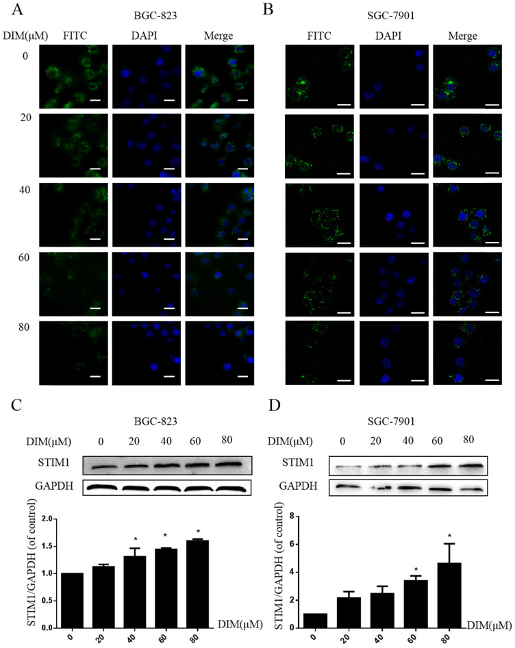
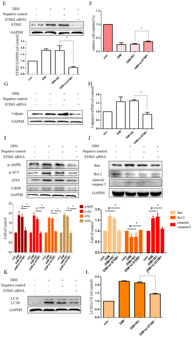
Figure 6.
Effect of SOCE on DIM-induced cell apoptosis and autophagy by p-AMPK-mediated ER stress in BGC-823 gastric cancer cells. (A) Cells were treated with different DIM for 24h, fluorescence intensity Ca2+ in ER store was measured by laser scanning confocal microscope. Scale bar: 15μm. (B) fluorescence intensity Ca2+ in ER store was measured by laser scanning confocal microscope in SGC-7901. Scale bar: 25μm. (C, D) Western blot analysis of the levels of STIM1 after treatment with different DIM (0μM, 20μM, 40μM, 60μM, 80μM) for 24h. (E) The knockdown efficiency of STIM1 was detected by Western blot in BGC-823 gastric cancer cells transfected with NC siRNA or STIM1 siRNA. (F) Cells were transfected with NC siRNA or STIM1 siRNA for 48h, followed by incubation with 80μM DIM for another 24h, cells viability was detected by the MTT assay. (G, H, I, J, K, L) The protein level of Calpain, p-AMPK, p-ACC, ATF6, CHOP, Bax, Bcl-2, cleaved-caspase 3, LC3II/LC3I was detected by Western blot in cells transfected with control siRNA or STIM1 siRNA. (Values represent as the mean±SD of three independent experiments (n=3)(*p<0.05 compared with the control group, #p<0.05 compared with the DIM group)).


