Fig. 6.
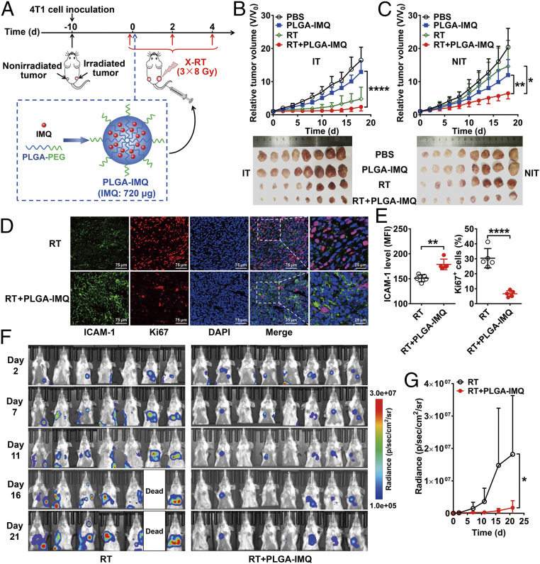
Systemic delivery of IMQ by PLGA nanoparticles enhances abscopal effect of RT in mice. (A) Schematic illustration of the structure of PLGA-IMQ and schedule of X-RT combined with PLGA-IMQ treatment. (B) Tumor growth curves of irradiated tumors (IT) in 4T1 tumor-bearing mice and photographs of tumors harvested at the endpoint (below) after the indicated treatments: control (PBS), PLGA-IMQ alone (PLGA-IMQ), RT alone (RT), and RT plus PLGA-IMQ (RT + PLGA-IMQ) (n = 8 per group). (C) Tumor growth curves of nonirradiated tumors (NIT) in 4T1 tumor-bearing mice and photographs of tumors harvested at the endpoint (below) after the indicated treatments. (D and E) Immunofluorescence staining (D) and quantification (E) of ICAM-1 and Ki67 in nonirradiated tumor tissues after treatment with RT or RT + PLGA-IMQ (n = 5 per group). (F and G) Serial bioluminescence images (F) of 4T1-fLuc tumor-bearing mice after the indicated treatments: RT alone (RT) and RT plus PLGA-IMQ (RT + PLGA-IMQ) (n = 7 to 8 per group). Quantitative results (G) are presented as quantified bioluminescence signals from the lungs. Numerical data are presented as mean ± SD. *P < 0.05; **P < 0.01; ****P < 0.0001 by unpaired Student t test (E) or two-way ANOVA (B, C, and G).

