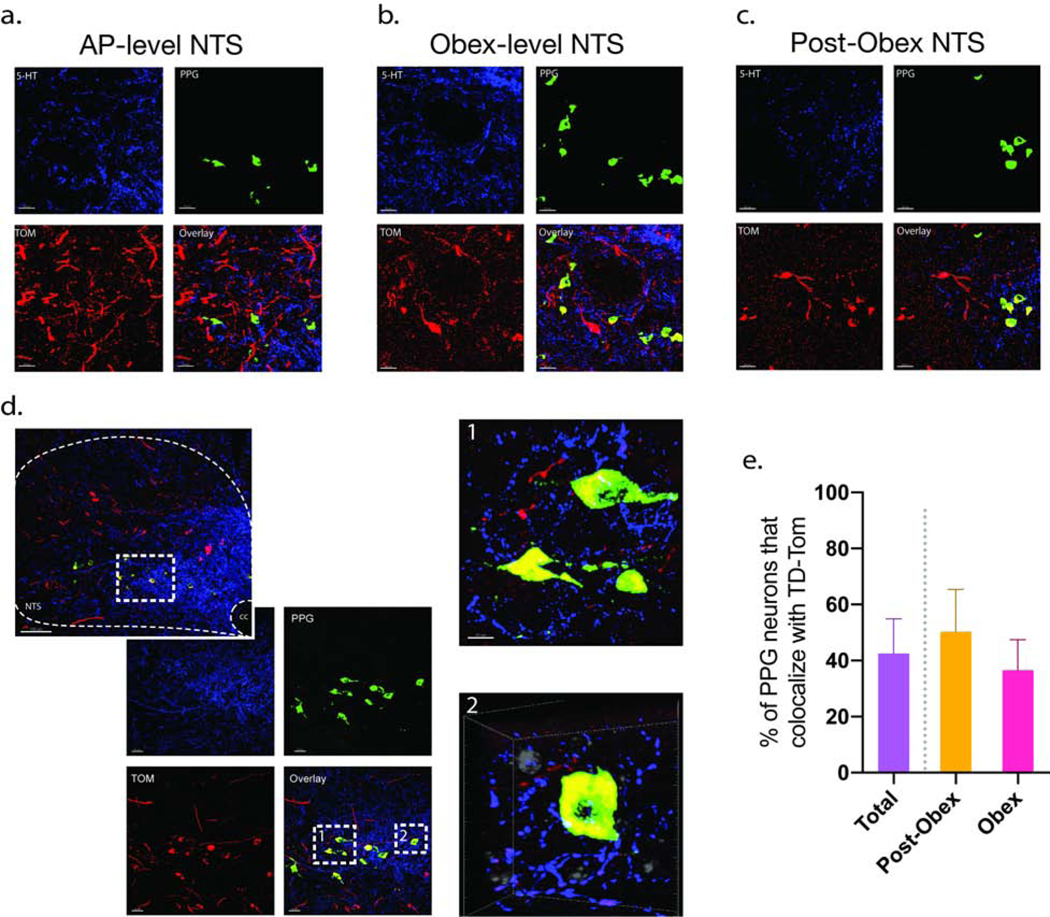
Fig 6. RMg neurons project mono-synaptically to PPG neurons in the NTS.
FISH/IHC was conducted on TD-Tom tagged NTS sections using an mRNA probe for PPG neurons (green) and a 5-HT antibody (blue) (n = 4). Neurons showing PPG/TD-Tom co-localization were identified in the AP- level NTS (a), Obex-level NTS (b), and Post-Obex NTS (c) sections. A representative obex-level NTS section at ×20 and x40 magnification is shown in (b), PPG are shown in green, TD-Tomato-expressing neurons in red, and 5-HT fibers in blue. Subpanel b-1 shows an optical zoom of two neurons co-localizing PPG and TD-Tom. Representative frame of three-dimensional rotational video showing PPG and TD-Tom co-expressing neurons in close opposition to 5-HT axons Subpanel b-2 (video can be found in the supplement data). The video was obtained from a z-stack collected from the mNTS at the level of the obex with the × 40 oil-immersion objective and a 4–5 optical zoom. A total of 43% of PPG neurons co-localized with TD-Tom, at the level of the pre-obex there was a 50% co-localization while at the obex-level there was a 37% co-localization (c).

