Figure 1.

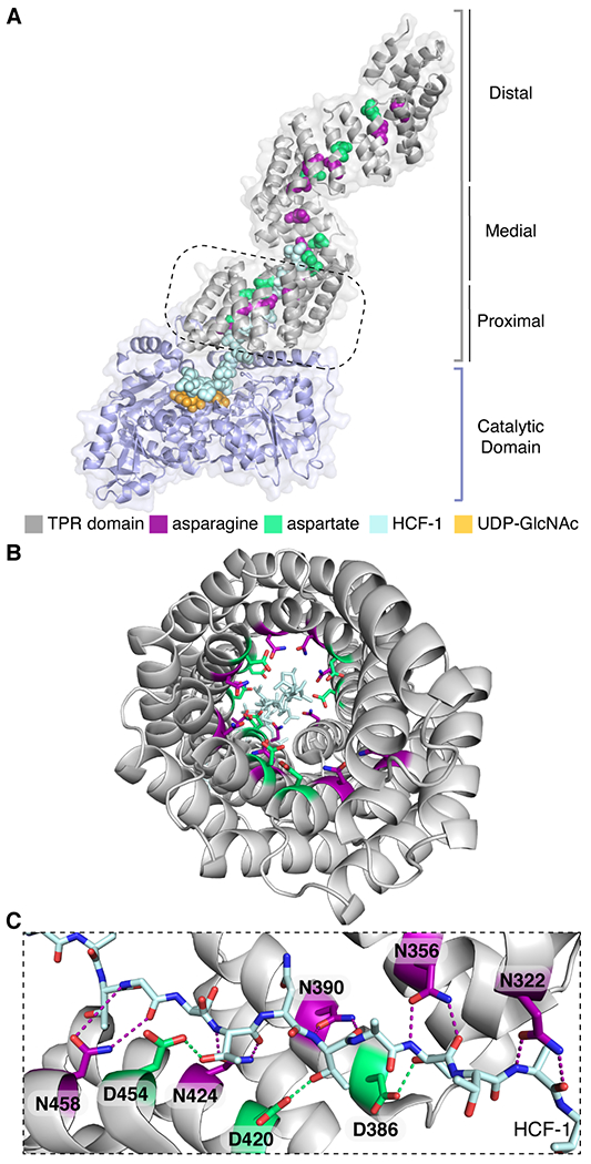
Conserved asparagine and aspartate ladders line the entire TPR lumen of OGT. (A) Composite structure (PDB 1W3B and 4N3B) of full-length OGT complexed with a peptide (HCF-1pro; light blue) that extends from the active site into the proximal TPR lumen (boxed region).18,24 The TPR lumen is lined by conserved asparagine (magenta) and aspartate (green) ladder motifs. (B) View through OGT’s TPR domain from the N-terminus. The peptide binds towards the C-terminal TPRs. (C) TPR asparagine side chains anchor the HCF-1pro peptide backbone; TPR aspartates contact polar groups on side chains of the bound peptide (the orientation shown is 90° from that in the dashed boxed in A).
