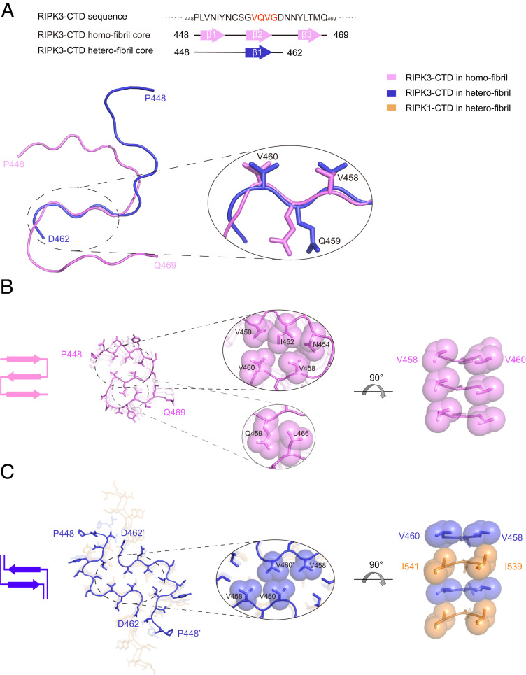
Fig. 5.
Structural comparison between RIPK3-CTD homofibril and heterofibril in complex with RIPK1-CTD. (A) The fibril core sequences of RIPK3-CTD homofibril and heterofibril are shown (Top). Overlay of one RIPK3-CTD subunit from RIPK3-CTD homofibril and heterofibril structures is shown (Bottom). The structure of the best-aligned VQVG is zoomed in. The rmsd of the four Cα atoms is 0.646 Å. (B) Top view of the RIPK3-CTD homofibril. The fibril contains one protofilament. The intramolecular interfaces within one RIPK3-CTD subunit are zoomed in. (C) Top view of the RIPK3-CTD heterofibril. The fibril contains two protofilaments. The intermolecular interface between two RIPK3-CTD subunits is zoomed in. The structures are shown in sticks. The sidechains in zoom-in views are shown in spheres. Topology of the fold in one layer is shown on the left.

