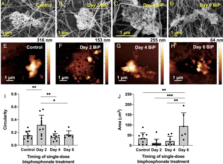
Fig. 6.
BiP treatment altered microcalcification morphology in a time-dependent manner. SEM (A–D) and AFM (E–H) were used to image microcalcifications formed in 3D collagen hydrogels that were treated at different times with a single dose of BiP. Microcalcification circularity (I) and size (J) significantly varied across time points (n = 4 biological replicates for control, day 2, day 4 groups, n = 2 biological replicates for day 6 group; *P < 0.05, **P < 0.01, ***P < 0.001, error bars represent SD).

