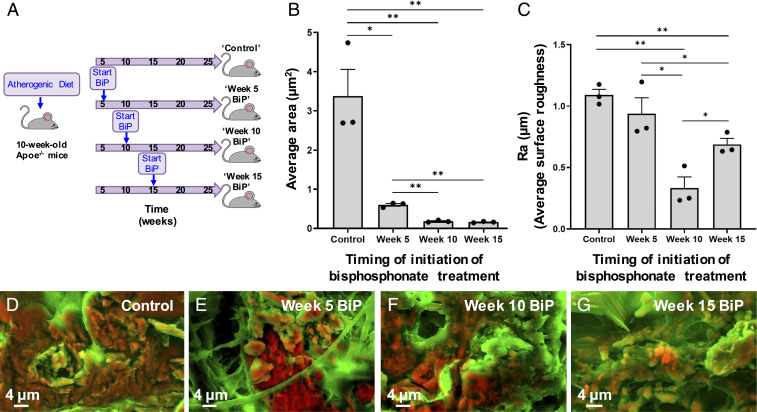
Fig. 8.
BiP treatment altered atherosclerotic plaque-associated calcification morphology in a time-dependent manner. ApoE−/− mice were fed an atherogenic diet and started on twice weekly BiP (ibandronate) treatment at various stages of atherosclerotic plaque development and calcification (A). Histological sections of aortic tissue were imaged using DDC-SEM. Quantitative image analysis was performed to measure the average area per individual calcification in each image (B) and the average surface roughness of calcific mineral in each image (C, n = 3 biological replicates per group; *P < 0.05, **P < 0.01, error bars represent SE of mean). Representative DDC-SEM images from each treatment group are shown (D–G).

