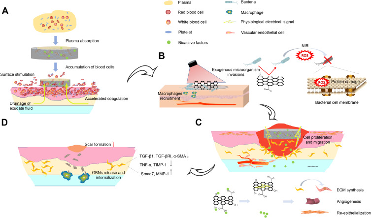
Figure 5.
Schematic illustration of effect and putative mechanisms of GBNs on wound healing in multiple wound healing stages in sequence. (A) In the hemostasis phase, the high specific surface area enables nanofibers to absorb plasma fast. The oxygen polar groups can instantly stimulate erythrocytes and platelets at the interface, further promoting blood coagulation. (B) The composite scaffolds exhibit good antibacterial performance owing to the small pore size to inhibit microorganism invasions and the photothermal properties of GBNs. (C) Released bioactive factors and GBNs act synergistically on fibroblasts, keratinocytes and endotheliocytes et al during proliferation phase. (D) The presence of GBNs inhibits the fibroblasts from overactivation, accompanied by the down-regulation of scar-related genes. Highly expressed MMP in the extracellular matrix could facilitate the macrophage to internalize the GBNs through endocytosis, leading to the biodegradation.

