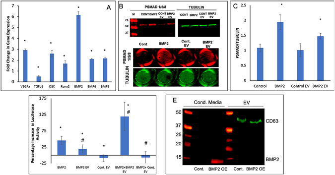
Figure 3: BMP2 FEEs potentiate the BMP2 signaling cascade:
A) Fold change in osteogenic gene expression (w.r.t untreated control) after HMSCs were treated with BMP2 EVs (1x10⁸ EVs per 250,000 cells) for 72 hrs. * Represents statistical significance w.r.t untreated control group (n=4, student’s t-test). B) Representative western blot (n=3) showing phosphorylated SMAD 1/5/8 (red lanes to the left) and tubulin (green to the right) after treatment of HMSCs with rhBMP2, Control EVs and BMP2 EVs. Note the increase in the band intensity for phosphorylated SMAD 1/5/8 after treatment with positive control BMP2 and with BMP2 EVs. For quantitation, an in-cell western was performed in 96 well plates (n=5). The image below the blots shows representative images of 96 well plates. C) Graphical quantitation of the intensity in each well for the in-cell western assay. *represents statistical significance w.r.t control as measured by Tukey’s ad-hoc pairwise test post ANOVA. Note that in line with the western blot data, the quantitative results show the phosphorylation of SMAD1/5/8 in the presence of BMP2 and BMP2 EVs. D) Graphical representation of SMAD 1/5 specific luciferase reporter assay that shows percentage increase in luciferase activity of the SMAD 1/5 specific reporter with the different treatments. Note the increase in activity after treatment with rhBMP2, BMP2 EVs and the combination of BMP2 and BMP2 EVs. * represents statistical significance w.r.t untreated control and # represents statistical significance w.r.t the rhBMP2 treated group (n=4 for all groups) as measured by Tukey’s ad-hoc pairwise test post ANOVA. E) Dual immunoblot for BMP2 (red) and CD63 (green) showing the presence of BMP2 in the EV-depleted conditioned medium from the BMP2 OE cells but not in the EV protein isolates of the control cell conditioned medium. CD63 was observed in the EV protein isolates only.

