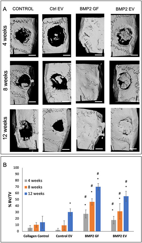
Figure 5: BMP2 EV mediated bone regeneration:
A) Representative μCT images showing regeneration of bone in 5mm calvarial defects that were treated with plain collagen sponge (Control), collagen sponge containing control EVs (Ctrl. EV, 5x10⁸ EVs per defect), collagen sponge containing BMP2 (BMP2 GF 5μg/defect) and collagen sponge containing BMP2 EV (5x10⁸ EVs per defect) at 4, 8- and 12-weeks post wounding. The arrow in the 12-week BMP2 GF group shows ectopic bone formation. Scale bar represents 2.5mm. B) Volumetric quantitation of the μCT data expressed as percentage bone volume regenerated with mineralized tissue (n=6 defects per group per time point). * represents statistical significance with respect to the collagen control group (no EV) and # represents statistical significance with respect to the control EV group. The significance was calculated using Tukey’s ad-hoc test following one-way ANOVA.

