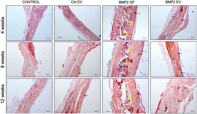
Figure 6: Histological evaluation of calvarial defects:
Images are representative light microscopy images of H&E stained demineralized calvarial samples of defects treated with plain collagen sponge (Control), collagen sponge containing control EVs (Ctrl. EV), collagen sponge containing BMP2 (BMP2 GF) and collagen sponge containing BMP2 EV after 4, 8- and 12-weeks post wounding. The black arrows in the images point to regenerated bone tissue. The yellow arrows in the BMP2 GF group point to fat deposits within the regenerated bone. Scale bar represents 200μm in all images.

