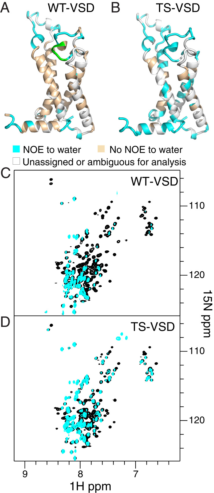
Fig. 3.
NMR reveals increased hydration within the water crevices of TS-VSD. Residues within WT-VSD (A) and TS-VSD (B), with visible crosspeaks to water in an 15N-edited NOESY-TROSY experiment are colored cyan on the homology model of the Shaker-VSD. Residues without crosspeaks to water resonances are colored wheat, and residues without assignments or peak overlap are colored gray. The raw data for WT-VSD (C) and TS-VSD (D) are shown below. The overlay of the 15N-1H TROSY-HSQC spectrum (black) and the water plane (∼4.58 ppm at 45 °C) from the 15N-edited NOESY-TROSY spectrum (cyan) clearly shows which backbone amide resonances have crosspeaks to water due to close proximity of water or H/D exchange. For WT-VSD (A and C), crosspeaks to water are not observed for the majority of residues on the transmembrane helices, while many more residues have crosspeaks to water in TS-VSD (B and D). This indicates increased water occupancy around the water crevices within the VSD. WT-VSD and TS-VSD are both stably embedded in the same way in the LPPG micelle (SI Appendix, Fig. S5), so the increased hydration of the water crevices in TS-VSD is not due to mutation-induced disruption of the protein/micelle complex. In addition, T265 to E268 in the S1–S2 linker of WT is colored green in A. Doubled resonances are observed for this region in the 15N-1H TROSY HSQC spectrum, but NOESY crosspeaks to water are only observed for one resonance, indicating that the two conformations have different water accessibility.

