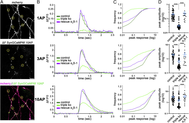
Fig. 2.
α2δ subunits are essential for activity-induced presynaptic calcium transients. (A) Putative synaptic varicosities from neurons cotransfected with SynGCaMP6f and mCherry were selected in Fiji/ImageJ using the ROI tool (yellow circles). For quantification of the presynaptic calcium transients, regions were transferred to the corresponding recordings of SynGCaMP6f fluorescence, shown here as fluorescence change by subtracting an averaged control image from an image averaged around the maximal response. (Scale bar, 10 µm.) (B) SynGCaMP6f fluorescence (ΔF/F0) for stimulations with 1 AP, 3 APs, and 10 APs at 50 Hz. Lines show the mean fluorescence traces for control (50 cells from three independent cultures), TKO/KD (triple KO, 50 cells from three independent cultures), and the rescue condition expressing α2δ-1 together with SynGCaMP6f and mCherry (19 cells from two independent cultures). (C) Cumulative frequency distribution histograms of peak fluorescent responses (ΔF/F0) from all recorded putative synaptic varicosities of α2δ TKO/KD (light green), double-heterozygous control (dark green), and α2δ-1 overexpressing TKO/KD neurons (rescue α2δ-1, purple) in response to stimulations with 1, 3, or 10 APs (number of synapses: control, 1,100; triple KO, 1,100; rescue α2δ-1, 418). (D) Quantification of peak fluorescent amplitudes in response to stimulations with 1, 3, or 10 APs. Each dot represents the mean of 22 synapses from one neuron [Kruskal–Wallis ANOVA with Dunn’s multiple comparison test: 1 AP: H(3, 119) = 81, P < 0.0001; 3 AP: H(3, 119) = 48, P < 0.0001; 10 AP: H(3, 119) = 26, P < 0.0001; post hoc test: ***P ≤ 0.001; 50 (control, triple KO) and 19 (rescue α2δ-1) cells from three and two independent culture preparations].

