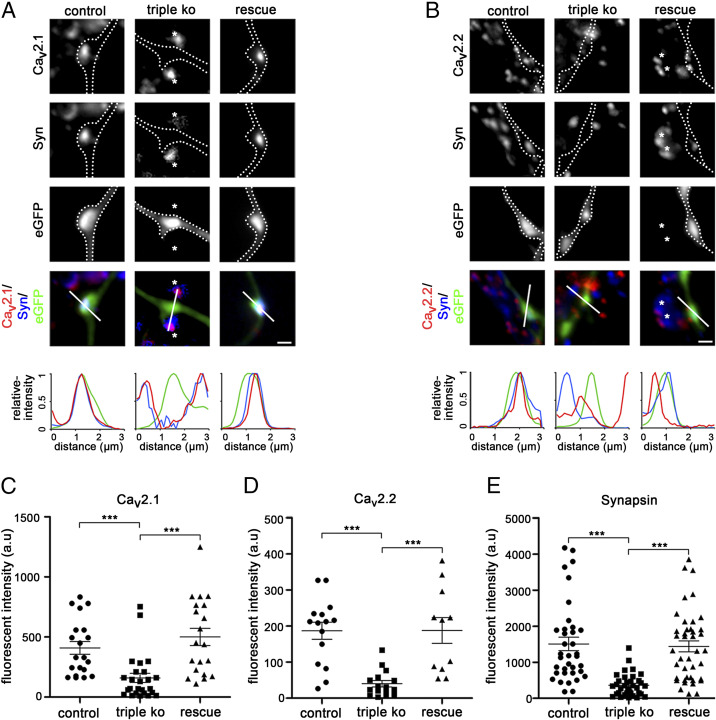
Fig. 3.
Failure of presynaptic calcium channel clustering and synapsin accumulation in α2δ subunit triple-knockout/knockdown neurons. (A and B) Immunofluoresence analysis of axonal varicosities from wild-type neurons (control, neurons transfected with eGFP only), TKO/KD neurons (triple KO, α2δ-2/-3 double-knockout neurons transfected with shRNA-α2δ-1 plus eGFP), and TKO/KD neurons expressing α2δ-2 (rescue, α2δ-2/-3 double-knockout neurons transfected with shRNA-α2δ-1 plus eGFP and α2δ-2). Putative presynaptic boutons were identified as eGFP-filled axonal varicosities along dendrites of untransfected neurons (SI Appendix, Fig. S4) and outlined with a dashed line. Immunolabeling revealed a failure in the clustering of presynaptic P/Q- (A, CaV2.1) and N-type (B, CaV2.2) channels as well as in the accumulation of presynaptic synapsin in varicosities from α2δ TKO/KD neurons (Middle). In contrast, wild-type control neurons (Left) displayed a clear colocalization of the calcium channel clusters with synapsin in the eGFP-filled boutons. The linescan patterns recorded along the indicated line support these observations. Note that the sole expression of α2δ-2 (Right) or the sole presence of α2δ-1 in synapses from neighboring α2δ-2/-3 double-knockout neurons (asterisks in A and B) suffices to fully rescue presynaptic calcium channel clustering and synapsin accumulation. (C–E) Quantification of the fluorescence intensities of presynaptic CaV2.1 (C), CaV2.2 (D), and synapsin (E) clustering in control, TKO/KD, and α2δ-2–expressing (rescue) TKO/KD neurons [ANOVA with Holm–Sidak post hoc test, ***P < 0.001; CaV2.1: F(2, 58) = 10.8, P < 0.001, 16 to 25 cells from four to six culture preparations; CaV2.2: F(2, 37) = 13.7, P < 0.001, 11 to 16, two to four; synapsin: F(2, 99) = 15.5, P < 0.001, 30 to 36, five to eight; horizontal lines represent means and error bars SEM]. (Scale bars, 1 µm.)

