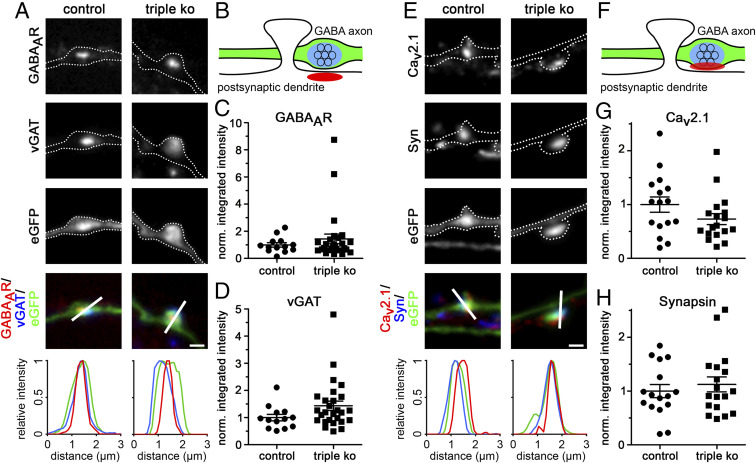
Fig. 5.
Presynaptic α2δ subunit triple-knockout/knockdown does not affect pre- and postsynaptic differentiation in GABAergic synapses. (A and E) Representative immunofluoresence micrographs of axonal varicosities from presynaptic α2δ-3 knockout (control) or TKO/KD (triple KO) cultured GABAergic MSNs. Transfected neurons (22 to 24 DIV) were immunolabeled for vGAT and the GABAAR (A) and CaV2.1 and synapsin (E). Colocalization of fluorescence signals within eGFP-filled axonal varicosities (axons are outlined with dashed lines) was analyzed using line scans. (B and F) Sketches depicting the expected staining patterns in A and E, respectively. (C, D, G, and H) Quantification of the respective fluorescence intensities in control and TKO/KD neurons (t test, GABAAR: t(38) = 0.8, P = 0.41, 13 to 27 cells from three culture preparations; vGAT: t(38) = 1.7, P = 0.10, 13 to 27 cells from three culture preparations; CaV2.1: t(32) = 1.6, P = 0.13, 16 to 18 cells from two culture preparations; synapsin: t(32) = 0.7, P = 0.51, 16 to 18 cells from two culture preparations). Values for individual cells (dots) and means (lines) ± SEM are shown. Values were normalized to control (α2δ-3 knockout) within each culture preparation. (Scale bars, 1 µm.)

