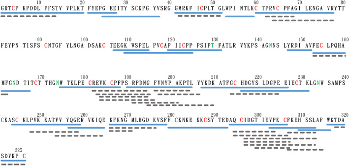
FIGURE 1.

Peptide map of the pepsin digested β2GPI. Native β2GPI was hydrolyzed by pepsin for 20 minutes. The peptides were then separated by HPLC and fragmented by tandem mass spectrometry. Sequences of peptides were identified by X!Tandem. The blue solid line is the peptide used in the subsequent structural presentation, and the gray dotted line is the sequence identified and analyzed, but not embedded in the protein structural presentation. The red 'C' indicates the Cys forming disulfide bond, and the green 'T' and 'N' indicate the glycosylated Thr and Asn
